【新增设备】Gatan Elsa冷冻传输样品杆,型号:698
2023-05-15 15:13 吴联翱Gatan Elsa冷冻传输样品杆,型号:698已于近日完成安装工作。Elsa™冷冻传输样品杆是全新一代单倾液氮样品杆,专为在液氮温度下将样品无霜转移至透射电镜(TEM)中而设计。 该样品杆主要用于在冷冻电镜(cryo-EM)中对辐照敏感的含水样品的单颗粒成像
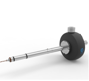
性能优点:
• 全新设计的杜瓦瓶,容量更大:液氮容量增加至原来的2.5倍
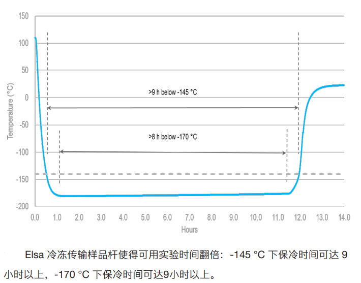
• 更长的保冷时间:提供长达8小时以上的稳定、高分辨率成像
• 更小漂移率,小于1.5 nm/min:确保在数据采集过程中高质量成像
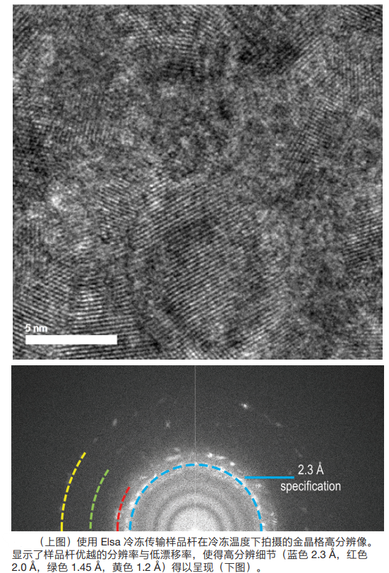
• 更高成像分辨率,优于2.3 Å:即使在冷冻条件下也能实现高分辨率成像
• 中心对称设计:消除样品杆倾转过程中的重心偏移,减小层析成像中样品杆稳定所需时间与漂移
应用领域:
• 冷冻电镜(Cryo-EM)
• 冷冻层析成像(Cryo-tomography)
• 电子晶体学
• 纳米颗粒成像
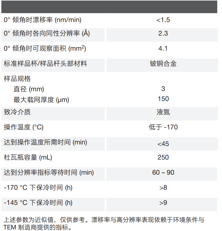
技术规格:
应用案例:
1.Frozen-hydrated rotavirus double-layered particles

Image courtesy of Dr. Debbie Kelly, Virginia Tech, Carilion
METHODS
frozen-hydrated rotavirus double-layered particles (0.01 mg/mL concentration, 2 µL volume)
prepared on Affinity Grids decorated with His-tagged protein A and antibodies against the outer capsid protein, VP6
specimen prepared using Cryoplunge™ 3 instrument with GentleBlot™ technology on C-flat™ 2/1 grids, Protochips, Inc.
specimens examined under low dose conditions at 120 kV using a 626 liquid nitrogen cryo transfer holder
recorded at 50,000x, dose of ~5 electrons/Å2
2.Frozen hydrated lipid vesicles
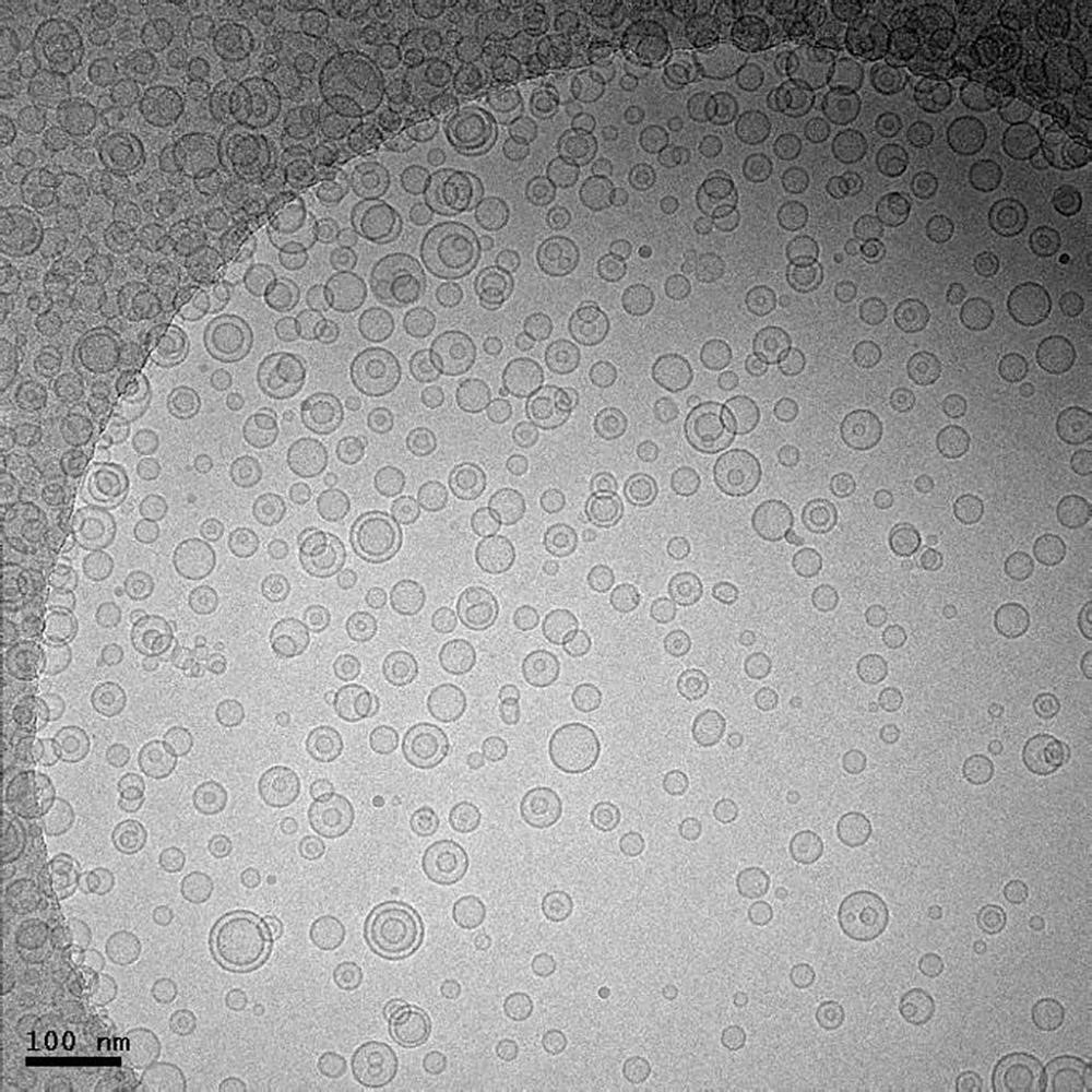
Image courtesy of Jessica Goodwin and Htet Khant, National Center for Macromolecular Imaging, Baylor College of Medicine, Houston, TX
METHODS
prepared using Cryoplunge™ 3 instrument
image was recorded using UltraScan 4000 camera
TEM magnification of 25 kx at 200 keV
electron dose of ~20 e-/Å2
910 multi-specimen single tilt cryo transfer holder
sample prepared on Quantifoil specimen support
used Solarus advanced plasma cleaner prior to freezing
3.Frozen-hydrated image of the Ndc80 complex decorated microtubules
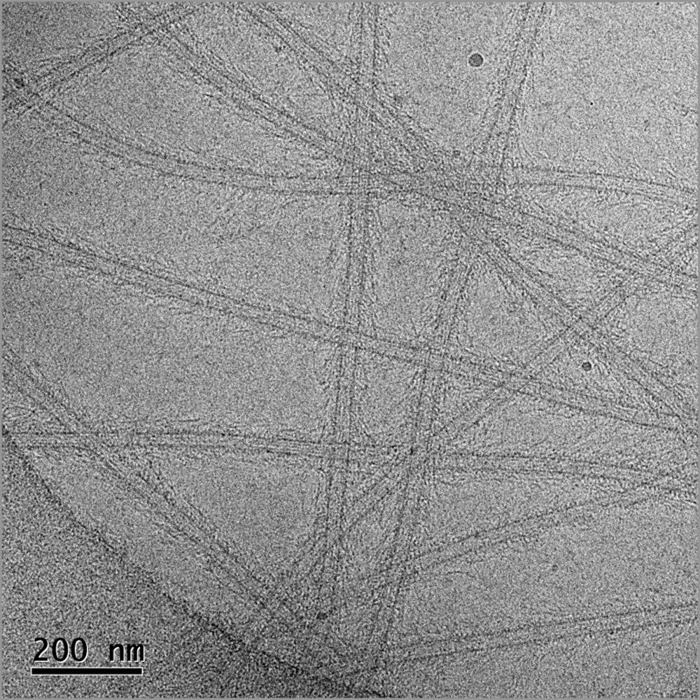
Image courtesy of Dr. Elizabeth Wilson‐Kubalek, Cell Biology, The Scripps Research Institute, La Jolla, CA
METHODS
magnification 30kx
electron dose 40 e‐/Å2
specimens prepared on CFlat™ Protochips, Inc. CF‐2/1‐4C‐50 holey carbon grids
plasma cleaned using the Solarus® advanced plasma cleaning system
frozen hydrated preparations produced using Cryoplunge™ 3 instrument
image recorded at 100 kV on a TEM equipped with a model 626 70° single tilt liquid nitrogen cryo-transfer holder
4.Lattice images of gold particles deposited on a Quantifoil carbon film
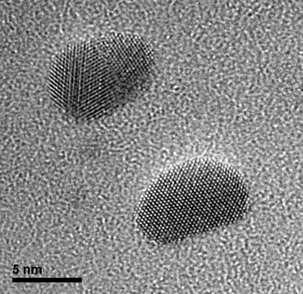
Image courtesy of Dr. John Berriman and Dr. Christian Kübel at KIT, Institute of Nanotechnology, Karlsruhe, Germany
Lattice images of gold particles deposited on a Quantifoil® carbon film with grid held at -176 °C using the 914 high tilt liquid nitrogen cryo transfer tomography holder.
METHODS
imaged using an UltraScan® 1000 2k x 2k camera
300 kV
8 s exposure
microscope magnification of 380 kx
5.Cryo-EM images of T7 phage
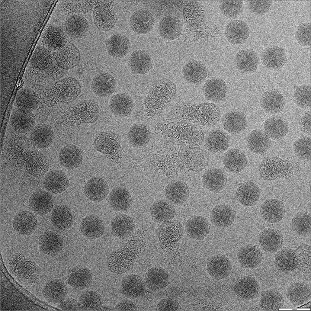
Image courtesy of Dr. Zheng Liu, Markey Center for Structural Biology, Department of Biological Sciences, Purdue University
Grid was prepared using Cryoplunge™ 3 instrument. Image was recorded at 300 keV using 626 liquid nitrogen cryo-transfer holder.
Scale bar 100 nm